
Back to:
Previews - click to zoom in
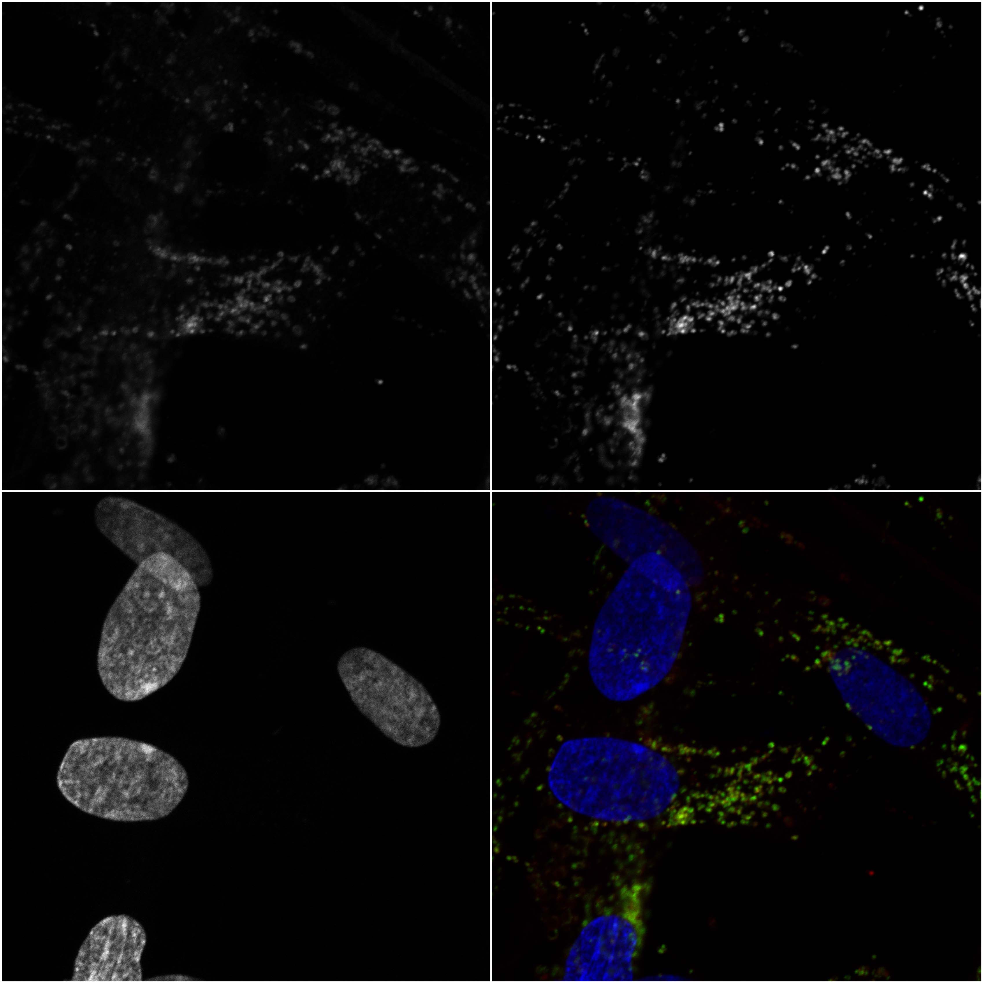 |
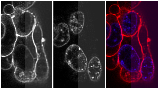
Selected lysosomal markers in human fibroblasts Human fibroblasts stained for cathepsin D (top left), beta glucocerebrosidase (top right), DAPI nuclear staining (bottom left) and merged image (bottom right). Plate was prepared in ImageJ sft. |
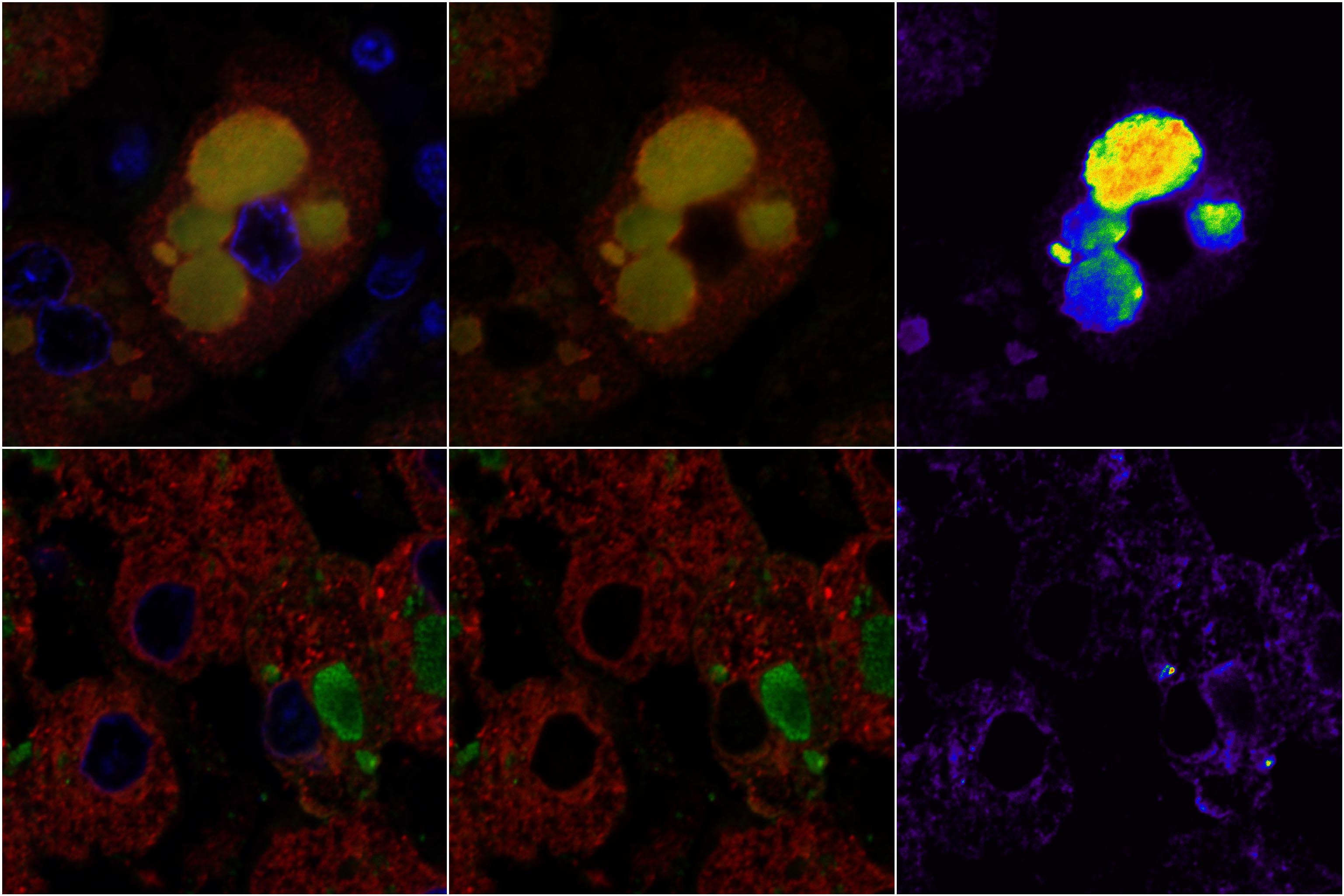 |
Fibrinogen deposits in two different cases of fibrinogen storage disease (liver tissue) Fibrinogen (green), protein disulfide isomerase (PDI) - endoplasmic reticulum marker (red), DAPI nuclear stain (blue). Rightmost images represent colocalization maps of red/green channels using overlap coefficient (0-1) scaled with appropriate LUT (calculated in Huygens sft). Plate was prepared in ImageJ sft. |
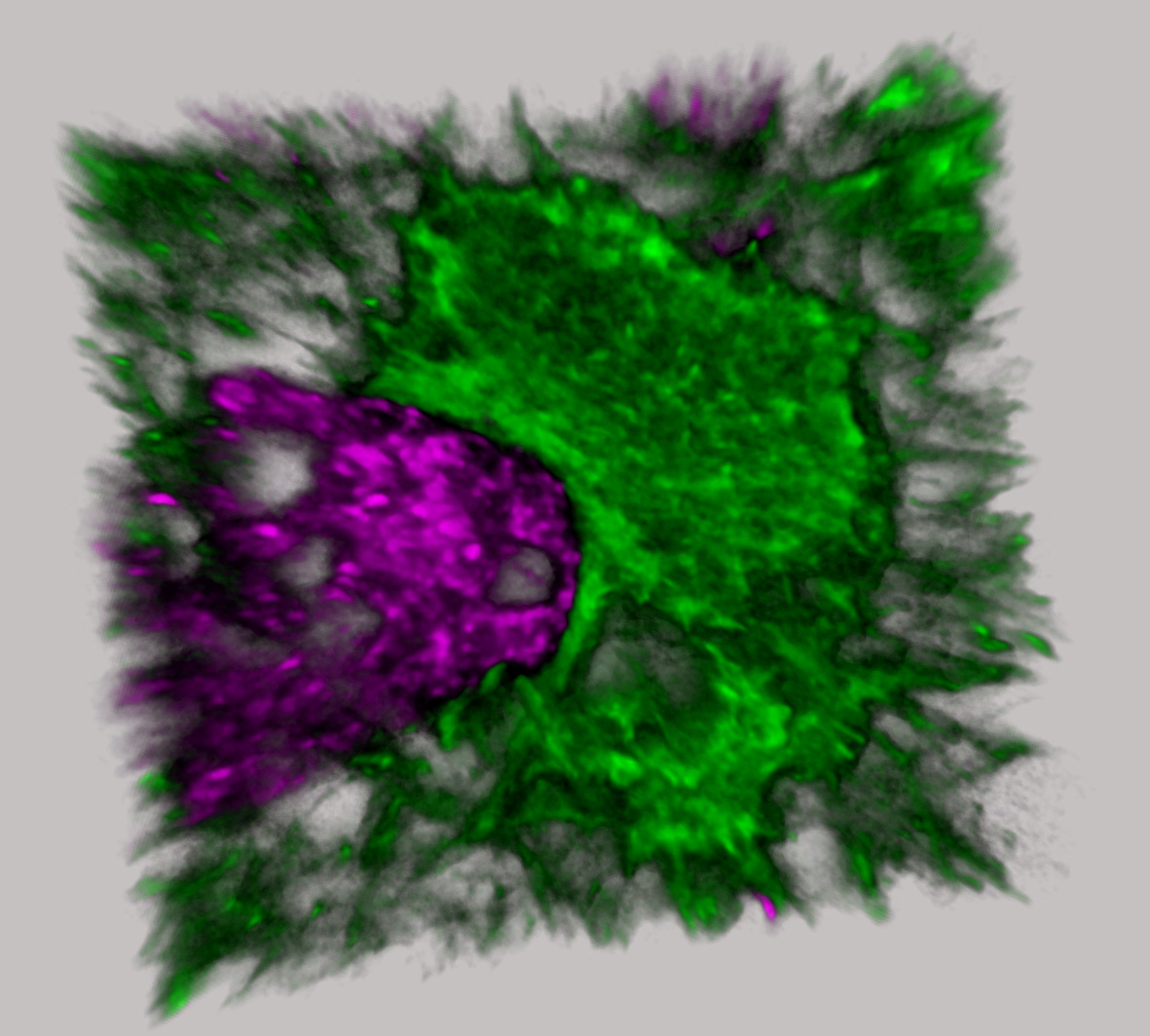 |
3D rendered image of glial phagocyte (violet) and astrocyte (green). Reconstruction performed in Imaris sft. |
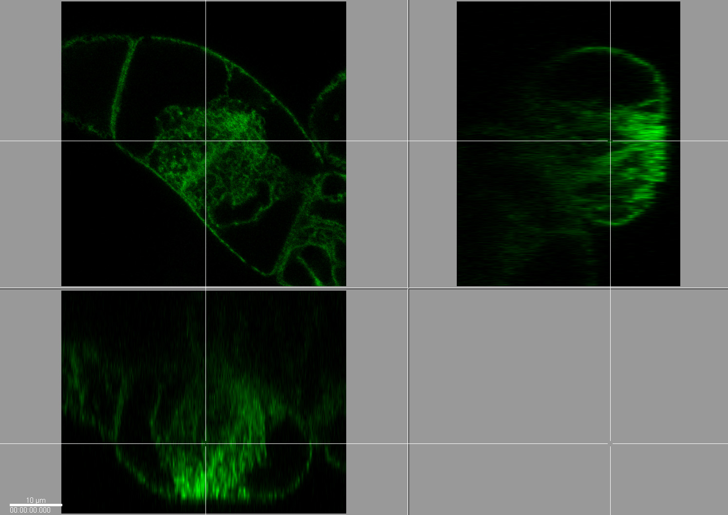 |
Tobacco expressing GFP in endoplasmic reticulum, orthogonal view (sample is courtesy of Dr.Petrasek, Institute of Experimental Botany Academy of Sciences, Czech Republic). |
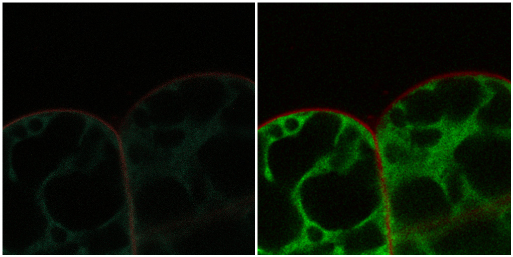 |
Tobacco expressing GFP in endoplasmic reticulum labeled with FM 1-43 (Molecular Probes - Invitrogen) for cell membrane. Image acquired in the spectral mode (left) of Nikon C1si. Right panel corresponds to spectrally unmixed image. Plate was prepared in ImageJ sft. |
 |
Att20 cells with DAPI labeled nuclei and FM 1-43 labeled cytoplasmic
membrane (Molecular Probes). Right side of the image was deconvolved with Huygens Professional software (SVI, Hilversum, The Netherlands) |
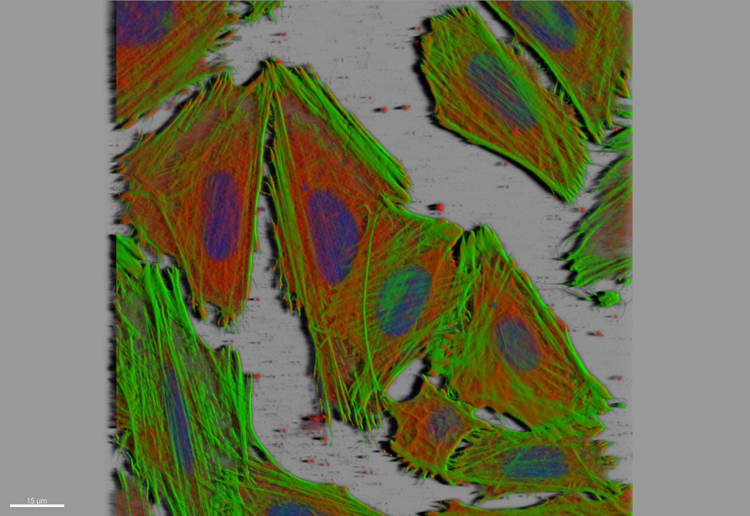 |
Actin cytoskeleton component (green), vinculin - adherent junctions
(red), nucleus - blue. Z-stack image rendered in Imaris Personal. |
 |
Secretory granules in cultured cells. Effect of deconvolution on wide-field volume images, the image acquisition was done on the TE2000 inverted microscope equipped with Vosskuhler CCD-1300 camera. The deconvolution was performed in Huygens Professional sft, image rendering was done in Imaris Personal. A,C - original data; B,D - deconvolved images. |