
Single-wall carbon nanotubes (SWCNT) - oxygen plasma treatment
The effect of the oxygen plasma treatment (1 min, 5 min and 30 min) of single-walled carbon nanotubes (SWCNTs) on their biocompatibility was investigated. The surface properties of SWCNTs after the oxygen plasma treatment were monitored by contact angle measurement, scanning electron microscopy and Raman spectroscopy. To test the biocompatibility of the oxygen plasma treated SWCNTs well characterized human osteoblastic cell line (SAOS-2) was used. Our results show that human osteoblasts cultivated on 5 min oxygen-plasma treated SWCNT films form confluent layer with pronounced focal adhesions under the whole cell body whereas they form a semiconfluent layer with focal adhesions distributed only marginally on 1 min oxygen-plasma treated SWCNT films as well as on pristine SWCNT films. Moreover, in case of 30 min oxygen-plasma treated SWCNT film, osteoblasts grow solitarily with very tiny focal adhesions on the periphery. Although 5 min oxygen-plasma treatment causes significant defects and slight change of SWCNTs morphology, from the cellular point of view it is the most convenient surface for osteoblasts massive adhesion and proliferation.
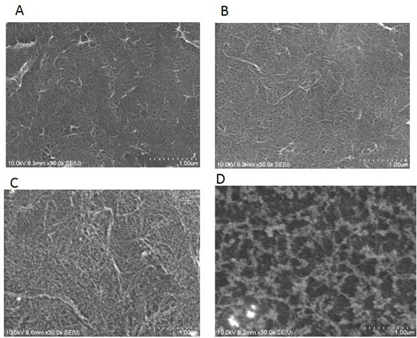
SEM images of pristine sample (A) and samples after oxygen-plasma treatment:
1 min (B), 5 min (C), 30 min (D). The scale bar is 1 µm
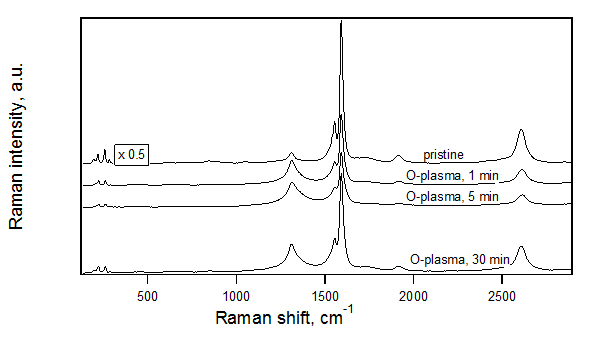
Raman spectra of pristine sample and samples after oxygen plasma treatment: 1 min,
5 min, 30 min (top to bottom). The Raman spectra were excited by 633 nm laser excitation energy.
The scale is same for all spectra except pristine sample which is scaled by factor 0.5.
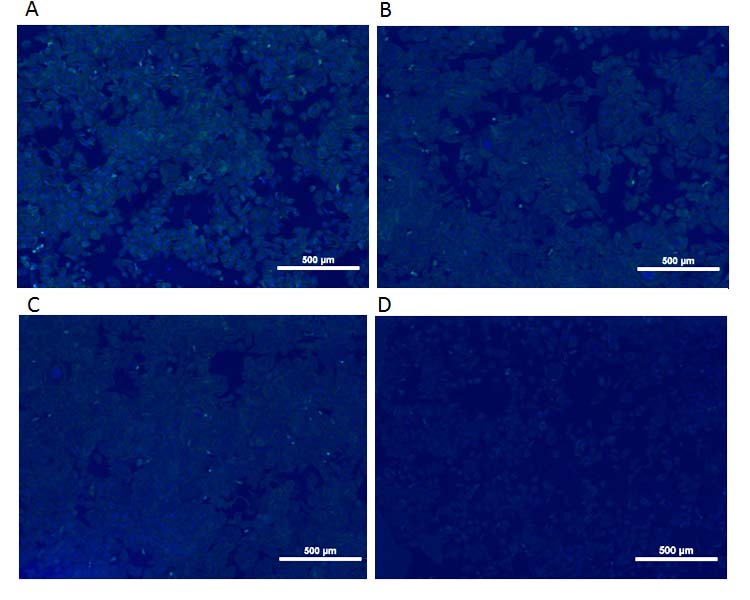 Fluorescent images of osteoblasts (SAOS-2) cultivated for 48 h on pristine sample
(A) and samples after oxygen plasma treatment: 1 min (B), 5 min (C), 30 min (D). The scale bar is
500 µm.
Fluorescent images of osteoblasts (SAOS-2) cultivated for 48 h on pristine sample
(A) and samples after oxygen plasma treatment: 1 min (B), 5 min (C), 30 min (D). The scale bar is
500 µm.
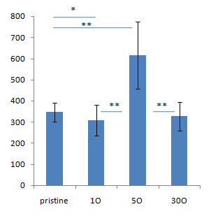
Cell number on different SWCNT types on area of 1mm2,*p<0.05,**p<0.01
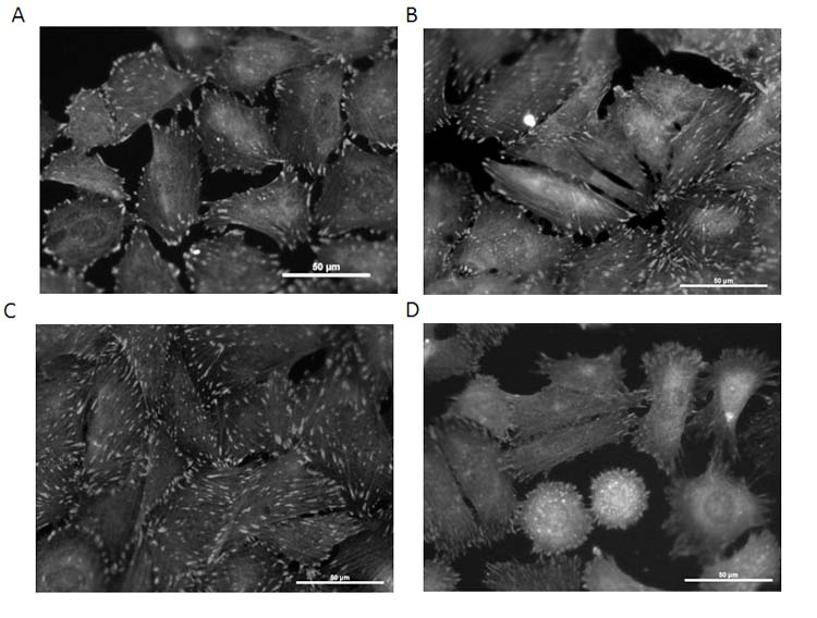
Fluorescent images of vinculin (structural focal adhesion protein)
in osteoblasts cultivated for 48 h on pristine sample (A) and samples after oxygen plasma treatment:
1 min (B), 5 min (C), 30 min (D). The scale bar is 50 µm.
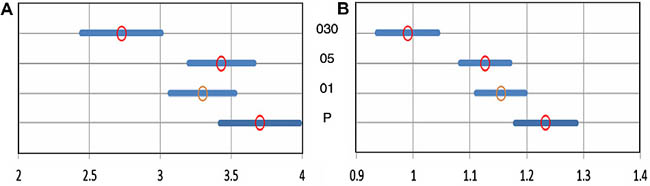
Estimated means of size of focal adhesions in cells on different
substrates and confidence interval for the difference between substrates. (A)
Long axis of focal adhesion and (B) short axis of focal adhesion, alpha = 0.01.
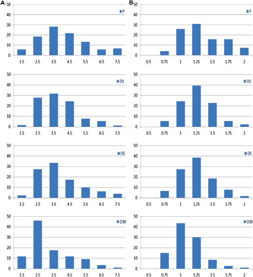
Relative frequency density histograms of size of focal adhesions in
cells on different substrates. (A) Long axis of focal adhesion and (B) short axis
of focal adhesion.