
| Rash Diseases and Skin InfectionsA Lecture Outline© Hanuš Rozsypal |
| Scarlet fever | Kawasaki syndrome | Toxic shock syndrome | Measles | Rubella | Roseola infantum | Megalerythema infectiosum | Other infections with maculopapular rash | Allergic eruptions | Chicken pox | Herpes zoster | Herpes simplex | Other infections with vesicular rash | Meningococcal disease | Other infections with petechial rash | Erythema multiforme | Erythema migrans | Erythema nodosum | Condylomata acuminata |
Thank you for your interest and I wish you a succesful exam of infectious diseases.
INTRODUCTION
Exanthematic infection = an infection where the rash is an obligatory, prominent, and fairly characteristic symptom. According to the morphology the rashes are classified usually to five (or more) groups
- maculopapular
- vesiculobullous
- petechial or purpuric
- erythematous
- nodular
 |
 |
 |
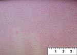
| Scarlatiniform rash (<1 mm) |
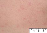
Rubelliform rash (1-3 mm) |
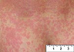
Morbilliform rash (3-5 mm) |
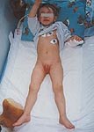
Incubation period: 2-7 days.
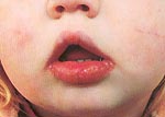
Symptoms and signs: Fever, tonsillitis, red strawberry tongue, generalized, fine, sandpapery rash.
 |
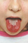 |
| Scarlet fever | Strawberry tongue with remnats of white fur |
| 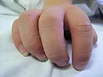 |
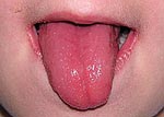
| Strawberry tongue | Šrámek´s sign - pale papules surrounding fingernails |
Differential diagnosis: Infection due to Arcanobacterium haemolyticum, Kawasaki syndrome, toxic-shock syndrome.
Therapy: Prokain-benzylpenicillin (50 000 IU/kg.day IM 3 days), then bezathinpenicillin (0,6 MIU IM), alternatives: fenoxymethyl-penicillin (PO), macrolids, 1st gen. cephalosporins.
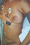
Etiology: Unknown, probably reaction to toxins (superantigens) produced by staphylococci or streptococci.
Pathology: Systemic vasculitis, in particular coronary arteriitis, which may lead to myocardial infarction and sudden death.
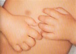
Symptoms and signs: Fever, conjunctivitis, rash, injected pharynx with dry, cracked lips and red strawberry tongue, swelling of the palms and soles with subsequent peeling of the skin. The illness may last 2-12 weeks!
Lab tests: leucocytosis, extreme thrombocytosis, high CRP. Echocardiogram: coronary aneurysms.
 |
 |
| Kawasaki syndrome | Kawasaki syndrome |
 |
 |
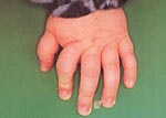 |
| Cracked red lips | Swelling of the hands | Peeling of the skin around the nails |
Pathogenesis: Some cases have been associated with the use of tampons during menstruation, other cases have followed staphylococcal infection at other sites.
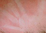 Dermografism blanch
Incubation period: 1-2 weeks (usually 9 days).
Symptoms and signs: Prodrome lasting several days, frequently consisting of fever (39-40°C), coryza, conjunctivitis, and cough. Koplik spots = white lesions on the buccal mucosa. Rash begins on the face 4 days after onset of symptoms. It spreads caudally over the next 3 days as the prodromal symptoms resolve. The rash lasts 4-6 days (it fades from the head downward). Desquamation may be present.
 |
 |
 |
| Koplik spots | Measles | Measles |
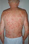
Incubation period: 12-23 days (average 18 days).
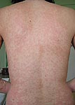
Symptoms and signs: Mild prodrome, mild catarrhal symptoms, fever is rarely high. On the first, second or third day, the maculopapular rash appears. It begins on the face and neck and rapidly spreads to the trunk and extremities. It fades without desquamation in 3-5 days. Adenopathy commonly affects occipital nodes. Arthralgias are common in young women.
 |
 |
| Rubella | Rubella |
Etiology: HHV-6, rarely HHV-7.
Incubation period: 1-2 weeks.
Symptoms and signs: Sudden onset, high fever that rapidly disappears on the 4th day. At this time, a rash that resembles rubella appears. The trunk is most affected.
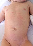 Exanthema subitum
Complications: Febrile cramps.
Incubation period: 1-2 weeks.
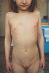
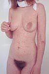
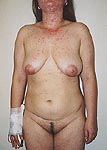
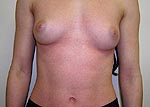
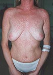
Symptoms and signs: Mild fever, sore throat, in many cases the prodrome is absent. Erythema on the face suggesting the marks of slapped cheeks, either concurrently or within a few days an annular and confluent erythematous rash emerges on the limbs and trunk. The rash usually resolves within a week, but can recur episodically for several weeks from exposure to heat (such as when bathing), cold, exercise, or stress.
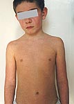 |
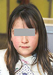 |
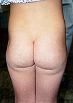 |
| Megalerythema infectiosum ("slapped cheeks" and rash on trunk) | Megalerythema infectiosum ("slapped cheeks") | Megalerythema infectiosum (rash on buttocks) |
 |
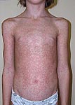 |
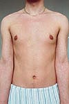
| Morbilliform rash | Morbilliform rash |
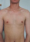
 |
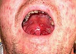 |
| Primary HIV infection - morbilliform rash | Primary HIV infection - aphthous ulcers in the mouth |
 |
 |
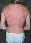 |
| Scarlatiniform rash | Morbilliform rash | Morbilliform rash |
|
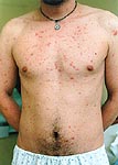
 |
 |
| Morbilliform rash | Erythematous exanthem | Morbilliform rash |
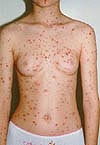
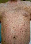
Incubation period: 10-23 days (average 17 days).
Symptoms and signs: Although there may be short prodrome of fever and malaise, rash is often the first evidence of illness. Laesions evolve in 5 stages: macules, papules, vesicles, pustulae, crusts. Typical laesion of the early stage is teardrop-shaped vesicle containing clear fluid, surrounded by reddened area (so called "glass pox"). Laesions appear in crops. Greatest concentration of the laesions occurs on the trunk, less on the face, extremities are generally less affected. Laesions in all stages may be found simultaneously. Most laesions are crusted by the 6th day. In many cases, the most irritating symptom is itching.
 |
 |
 |
| Chickenpox | Chickenpox | Secondary infection of chickenpox (varicella impetiginisata) |
Pathogenesis: Reactivation of VZV lying dormant in cells of a dorsal root or geniculate ganglion.
Symptoms and signs: An attack of shingles begins with pain and hyperaesthesia. Within a few days a cluster of vesicles appears in an area of a sensory nerve root.
 Herpes zoster faciei
Sequelae: Postherpetic neuralgia.
Therapy: Aciclovir.
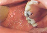
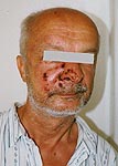
Symptoms and signs: A cluster of vesicles on an erythematous base that progresses to mucocutaneous ulcerations. The common sites are lips, facial skin, nailbed etc. Oral herpes:
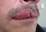 Oral herpes
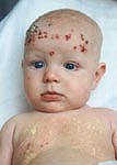 Eczema herpeticum (Kaposi´s varicelliform eruption)
 |
 |
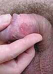
| Genital herpes in a man | Genital herpes in a woman |
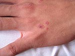
 |
 |
| Hand-foot-and-mouth disease | Hand-foot-and-mouth disease |
 |
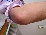
 |
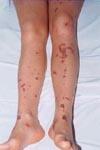 |
| Petechial eruption during meningococcaemia | Petechial eruption during meningococcaemia | Ecchymoses during meningococcal sepsis (>3 mm) |
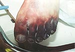 Gangrene of feet due to arterial thrombosis
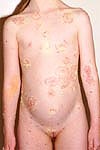
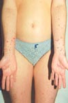
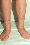
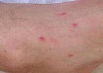
Symptoms and signs: Laesions begin as macules and papules of dark red to brown colour, may develop into blisters and are symetrically distributed on the trunk and extremities. Predilection sites are knees, elbows, palms, and soles. Mucosal involvement is ususally present and it is painful. The severe form with mucosal involvement is called Stevens-Johnson´s syndrome.
 |
 |
 |
| Laesions on elbows | Laesions on hands | Laesions on knees |
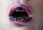 |
 |
| Inflammed and crusted lips | Laesions on trunk |
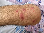
Etiology: Borrelia burgdorferi, particularly B. afzelii.
Incubation period: 3-32 days (after the tick bite).
Symptoms and signs: Painless flat erythematous lesion with partial central clearing usually localized on the thigh, groin, axilla etc. The diameter is several centimeter. It may be present for days to several weeks.
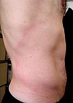 Erythema migrans
Pathogenesis: Sensitisation to a number of agents.
Symptoms and signs: Tender nodular lesions on the shins.
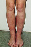 Erythema nodosum
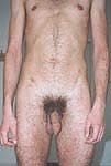
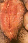
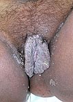
Symptoms and signs: Papules with irregular, verrucous surface, located around both the external genitalia or the anus.
 |
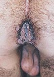 |
| Gigantic genital warts | Perianal warts |
References
- Džupová O. Exanthem of infectious origin. In: Hobstová J et al. Infectious Diseases. 1st ed. Karolinum, Prague 2003, pp.125-137
- Emond RTD, Rowland HAK, Welsby PD. Color Atlas of Infectious Diseases. 3rd ed. Mosby-Wolfe, Times Mirror International Publishers Limited 1995
The page was last updated 13-May-2004