Laboratorní vybavení
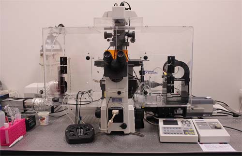
• Laser scanning confocal microscope (Nikon Eclipse TE2000 (inverted): two confocal heads – CSi with spectral detector and SFC (swept-field confocal), 4 lasers (405 nm, 488 nm, 561nm and 647 nm), Perfect Focus System for stabilization of Z table, set of different objectives, thermal and CO2 incubation devices, anti-vibration table (Melles Griot)
Confocal microscope Leica SP8X - freely selectable excitation laser line in range 470-670nm of continuous wavelength pulse laser combined with diode laser 405nm and higher power multi line argon laser, tunable acustooptical beamsplitter instead of standard dichroit, freely selectable emission signal (dispersion with glass prism), two high sensitivity hybrid detectors (time gating of the signal effectively filtering reflected/nonfluorescence signal), two photomultiplier tube detectors. The whole system is prepared for long term live cell experiments in controlled CO2/O2 atmosphere and temperature (cage incubator, automatic focus correction, DIC and PH objectives, and oil immersion objectives). Rapid cellular events can be recorded by 8 kHz resonant scanner. http://www1.lf1.cuni.cz/udmp/Microscopy/index.php
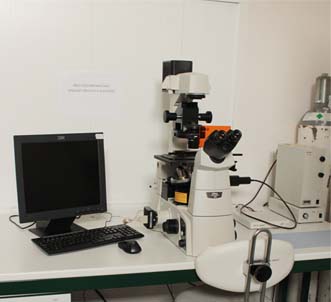
• Epifluorescence microscope – Nikon ETi-S and Nikon Eclipse E400 with set of objectives (phase contrast, long working distance), high sensitivity CCD cameras (DS-Qi1Mc, DS-U1), Intensilight.
• Image analysis software - NIS Elements, Huygens Professional Software, Imaris, ImageJ
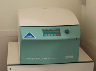
• Ultracentrifuge –Beckman-Coulter Optima L-90K
• Molecular biology and biochemical laboratory is equipped with standard facilities for isolation of nucleic acids and their manipulation and analysis – cloning, PCR, quantitative RT-PCR, sequencing (sequenators - SOLID4 System and capillary ABI 3500XL Avant Genetic Analyser, Life Technologies), gene expression and analysis (in prokaryotic and eukaryotic system) and work with proteins – 1D/2D protein electrophoresis, multiformate plate reader (Synergy 2, Biotek).
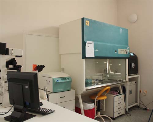
• Tissue culture laboratory is equipped with standard facilities as laminar flow boxes, CO2 incubator, Dewar bottle for sample storage in liquid nitrogen, epifluorescence microscope with CCD camera (Nikon E Ti-S)
Cells
SAOS-2 - osteoblast-like osteosarcoma cell lineHeLa G - cervical carcinoma cell line
MSC - mesenchymal stem cells (from "patients" suspected for haematological disease, but being negative)
Laboratory
