

Last updated: April 22, 2003
HTML validace NetMechanic:

PACS - GI images of unusual, atypical, misdiagnosed cases.
Individual pictures (images) could be enlarged (zoom) clicking on selected picture. Documentation presented on these pages has been digitalized on-line by using the GastroBase-II module (Information System of the Medical Clinics of Fac.Hospital, Prague). All data are presented anonymously with agreement of the directory of our Hospital and are intendt for postgraduate education of gastroenterologists.
One of the international resources could be mentioned the ELCANO project - Virtual Electronic Library of Unusual Clinical Cases, mainly in the gastroenterology. The URL address: www.imim.es/elcano/.
 GastroBase-II Information System with PACS
 Database of endoscopic/X-Ray/UltraSound images !!! NEW !!!
Cases:
 Leiomyoma, leiomyosarcoma, EUS images, included in the ELCANO library
 Leiomyoblastoma, hamartoma, EUS images
 Giant pancreaticolithiasis, Xray, ENDO images, included in the ELCANO library
 Endoscopic treatment of large adenoma of papila Vateri, ENDO, EUS images
 Endoscopic treatment of Zenker's diverticulum by argon coagulation, ENDO images

Case report No.1: Leiomyoma - leiomyosarcoma
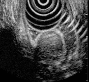
20yrs old female, presented in 1994 by dyspepsia, suspected intramural process was found on the gastroscopic examination. Endosonography was made in 1995 and a mass 25mm in diameter, with regular margins, originated from the 4th layer was found. This result was evaluated as leiomyoma and this diagnose was confirmed by aimed aspiration biopsy (EUS image No.1.1).
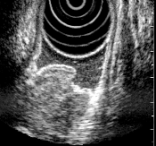
Repeated endosonography was made in 1996, extent of the mass was not changed, but inhomogeneity occured with a small ulceration on the top (EUS image No.1.2). Due to these facts a surgery has been recommended.
Leiomyosarcoma with liver and node metastatic process was found by operation. Conclusions: On this case could be demonstrateted impossibility to determined the degree of malignancy by EUS alone.
Case report No. 2: Leiomyoblastoma - hamartoma
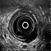
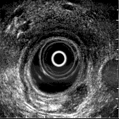
61 yrs old man with polypoid leasion of gastric wall (antrum, superficially exulcerated).
Endosonographic diagnosis (in the 1994) was leiomyoma, originated from the 4th layer (EUS image No.2.1), but due to inhomogeneity leiomyoblastoma was also possible (EUS image No.2.2). Aspiration biopsy with cytologic examination resulted tumorous cells, probably malignant. Operation was recommended and carcinoma-like tumor was proved by surgeon. The histology of this tumor: hamartoma containing various different tissues.
Case report No. 3: Giant pancreaticolithiasis
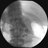
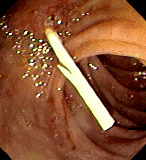
43yrs old male patient with cholecystectomy in 1974, chronic pancreatitis of alcohol etiology diagnosed in 1993. ERCP in 1993 showed giant pancreaticolithiasis of unusual extent (X-ray image No.3.1) - several stones with a diameter > 20 mm in dilatated ductus Wirsungi. Due to the size of stones an endoscopic lithotrypsy wasn't successful and so this situation is temporaly solved by repeated replacement of pancreatic stent (ENDO image No.3.2) every 6 months.
The patient is now without any troubles even that X-ray picture is not changing.
Case report No. 4: Endoscopic treatment of large adenoma of papila Vateri
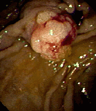
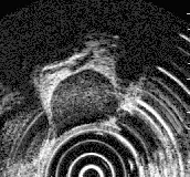
73yrs female patient presented in 1995 with heavy cholestasis. ERCP showed a large, multilobular, papila Vateri with diameter more than 40 mm (ENDO image No.4.1). Histology of the biopsy sample resulted the benigne adenoma. On the EUS picture papila was homogenous, with sharp margins, without signs of invasion into the duodenal wall (EUS image No.4.2). The situation has been solved by endoscopic resection.
Case report No. 5: Endoscopic treatment of Zenker's diverticulum by argon plasma coagulation
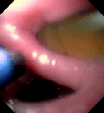
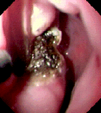
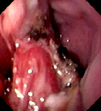
75yrs female patient presented in 1995 with "upper" dysphagia, pressure in jugular area after meal with repeated regurgitation of swallowed boluses. Oesophageal X-ray and endoscopy in 1998 showed the hypopharyngeal Zenker's diverticulum, the depth of which was almost 5 cm.
With regard to the patient's complaints we decided on the endoscopic treatment using the argon plasma coagulation (APC). Firstly, we performed standard upper endoscopy and introduced the nasogastric tube over the guide wire to make the tissue bridge between the oesophagus and the Zenker's diverticulum more pronounced (ENDO image No.5.1). Afterwards, we coagulated carefuly the bridge using the argon plasma beamer in two sessions with the interval of one day between the procedures (ENDO image No.5.2.). We achieved the distinct destruction of the tissue bridge with disclosing the cricopharyngeal muscle fibres on the top of the Zenker's diverticulum bridge (ENDO image No.5.3). Described treatment was successful and we didn't record any complications associated with the therapy.
The actual stage of this page  is running.
 The Czech and International Internet resources linked with this WWW page:


published on the server 1st Medical Faculty of Charles University, Prague.
Icons for Webmasters ::


Number of accesses to this page since August 1, 1999:

WebMaster: Petr Kocna -
kocna@mbox.cesnet.cz
HomePage: medical oriented or church and theology oriented


![]()
 GastroBase-II Information System with PACS
 Database of endoscopic/X-Ray/UltraSound images !!! NEW !!!
 Leiomyoma, leiomyosarcoma, EUS images, included in the ELCANO library
  Leiomyoblastoma, hamartoma, EUS images
 Giant pancreaticolithiasis, Xray, ENDO images, included in the ELCANO library
 Endoscopic treatment of large adenoma of papila Vateri, ENDO, EUS images
 Endoscopic treatment of Zenker's diverticulum by argon coagulation, ENDO images


 is running.
 The Czech and International Internet resources linked with this WWW page:


![]()


